
Last Updated on: 27th December 2025, 08:44 am
Very rarely, dentigerous cysts occur in the first decade of life given that most develop during adulthood in approximately the second to third decade of life.
Cysts of dental origin are benign and always involve a tooth that cannot complete its eruption or development process. However, only the crown is involved with the formation of cysts. The sac that contains the tooth inside the bone can accumulate fluid between the layers of enamel that surround the crown is not involved.
Dentigerous cysts develop mainly in the mandible; a low percentage are found in the upper jaw. The wisdom teeth are most associated with the development of cysts. Being benign, they do not endanger the life of the person, but they can cause serious damage if their growth is extensive. According to the ADA (American Dental Association), cysts can cause damage to the root of neighboring teeth and the bone that surrounds the tooth.
What is Dentigerous Cyst and what are there symptoms?
Generally, dentigerous cysts are painless and asymptomatic; however, their growth can affect other dental structures, causing:
• Tooth sensitivity
• Swelling
• Displacement of the teeth
• A bump at the site of the eruption
Small cysts do not have symptoms, and a diagnosis must be supported by an X-ray. Dentigerous cysts that generate some type of symptomatology must measure more than 2 centimeters in diameter.
How are Dentigerous Cysts Found?
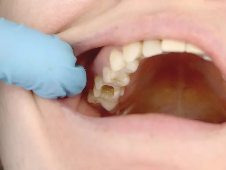
Dentigerous cysts are characterized by slow growth. They are often found in the back of the mouth and are associated with the development and eruption of the third molars or wisdom teeth. If the cyst is small, the patient may never become aware of its presence.
In many cases, dentigerous cysts are diagnosed through X-rays done during routine consultations.
What Causes Dentigerous Cysts?
Dentigerous cysts occur from the accumulation of fluid on top of teeth in the process of erupting but have not yet emerged. In general, these teeth lose their eruption strength and become trapped within the bone.
The appearance has no apparent origin; however, odontogenic cysts are related to genetic syndromes such as nevoid basal cell carcinoma syndrome, where the body lacks the gene that suppresses tumor growth. This type of syndrome is unfortunately also related to the development of cancers.
The most well-known theory of the development of dentigerous cysts states that the cyst originates after the crown of the tooth has been completely formed, through the accumulation of liquid between the enamel layers that surround it, their studies suggest that the formation of cysts can occur at any stage of tooth development. What all theories agree on is that they form when the teeth have not yet erupted.
The dentigerous cyst will always be associated with the crown of the permanent tooth, which is retained, i.e., not erupted. Very rarely it will occur in primary teeth. It commonly occurs in males between the second and third decade of life in a higher percentage in the lower jaw (70%-75%).
What do Dentigerous Cysts look like?
As mentioned, dentigerous cysts are asymptomatic; however, when the cysts are of a considerable size, generally greater than 2 centimeters in diameter, they can be observed clinically as they produce an expansion in the bone, giving the appearance of swelling on the face. It can feel hard and does not cause pain in the patient.

If the size of the cyst is very large, it can cause jaw fractures. For the patient, it can be painful without being unbearable, which is why its diagnosis is so difficult.
Generally, the diagnosis of these cysts is supported by a panoramic X-ray, since with this type of X-ray, the mouth and all neighboring structures can be observed.
Diagnosis and Treatment of Dentigerous Cysts
For the diagnosis of cysts, the dentist relies on an x-ray. A cyst is observed as a small translucent spot, and after diagnosis, the procedure will depend upon its size. If the cyst is small, the decision could be to leave it and carry out radiographic controls. But, if the size is large, the decision is the resection or removal of the cyst, a procedure called marsupialization.
How are Dentigerous Cysts Removed?
The removal procedure is performed under local anesthesia, while in some cases, the surgical approach must be done under general anesthesia, especially for larger cysts. The technique consists of making a cut that exposes the cyst; then once exposed, the tooth involved is removed and the fluid that surrounds it is drained with specialized instruments.
In some cases when the cyst is large, not only is the cyst removed but also the teeth are affected by its growth.
If the cyst is small, an incision can be made that allows only the fluid to be drained to help in the future eruption of the tooth. However, this surgical technique is not widely used since the diagnosis of dentigerous cysts occurs when it affects neighboring structures.
Other treatment options are:
• Jaw reconstruction surgery
• Comprehensive treatment between speech therapy, nutrition, and swallowing
Treatment follow-up must be done for life since large cysts can affect the surrounding bone of the tooth.
Possible Complications of Dentigerous Cysts
Removal of dentigerous cysts, even small ones, can prevent the following complications:
1. Infections: An infected cyst can cause periodontal and periapical infections in the tissues surrounding the tooth.
2. Loss of teeth: A cyst that goes untreated weakens the gums and tissues that surround it, causing the weakening of the neighboring teeth.
3. Fracture: Generally, the growth of the cysts can affect the bone, causing a fracture of the jaw.
4. Ameloblastoma: These rare tumors mainly affect the jawbone. If left untreated, the swelling can become cancerous, and in the worst cases, the disease can spread to the lungs and lymph nodes.
To avoid affectation, it is best to go to the dentist regularly. A timely diagnosis can avoid serious sequelae in the patient, especially the aesthetics and function of the teeth, thereby affecting quality of life.