
Last Updated on: 27th December 2025, 05:03 am
Hypomineralization of enamel, hypomineralization, or incisor-molar hypomineralization (IMH) is a genetic alteration that occurs when teeth are forming during the third trimester of pregnancy, in the primary teeth, or during the first three years of life in permanent teeth. Enamel hypomineralization consists of a decrease in the mineral content of the teeth.
This can be observed as white or yellowish spots that affect both function and dental aesthetics. The condition can occur after the teeth have erupted from the gums, but the problem is that the teeth are completely unprotected and can easily suffer from cavities, softening, cracks, and fractures.
It is estimated that hypomineralization occurs in 1 in 5 children in their primary or permanent teeth.
Why Does Hypomineralization of Enamel Occur?
The causes of hypomineralization are still unclear; however, recent studies show that this condition may be due to genetic predisposition. Other studies show that patients suffering from this condition are affected by external factors such as poor nutrition or excess fluoride at certain times of tooth development. Other factors are:
• Maternal smoking during pregnancy
• Vitamin D deficiency
• Part premature
• Lack of oxygen at birth or respiratory problems during the first years of life
• Fever
• Low birth weight
For a long time, it was believed that the use of antibiotics in the first years of life was a predisposing factor; however, the studies conducted to prove this theory are not conclusive. It is necessary to clarify that this condition is not the result of lifestyle practices such as diet or oral hygiene.
One of the factors that can exacerbate the effects of hypomineralization of enamel is acid exposure from sources like acidic foods, beverages, or stomach acids that lead to enamel erosion.
What Does Incisor-molar Hypomineralization Look Like?

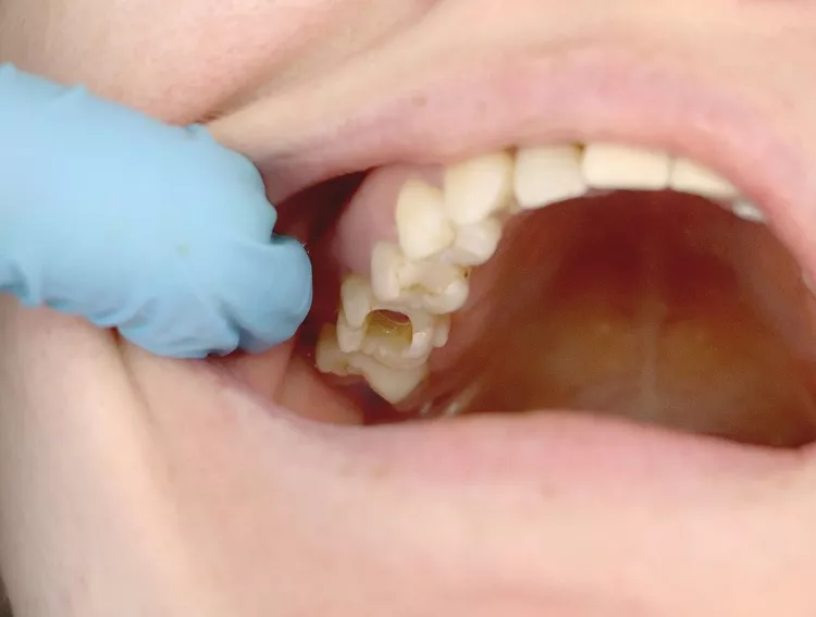
Hypomineralization usually occurs in the first molars and incisors (hence its name). The appearance of this condition can vary from white to yellowish and brown spots. Hypomineralization lesions are classified as:
• Light: Colorful defects (white spots) without apparent damage to the structure; generally only the biting surface of the tooth or molar is affected.
• Moderate: In this defect, yellowish or brown spots are observed on the bite surface and the cusps or reliefs of the teeth. These areas are also very susceptible to rupture due to the weakening of their structure. At this point, the tooth is also very susceptible to sensitivity.
• Grave: More extensive brown and/or yellowish spots are observed on the surface of the teeth or molars, and fractures and dental sensitivity are more frequent.
Is Hypomineralization of Enamel Painful?
Tooth sensitivity is a common symptom of this condition; however, it is a manageable discomfort depending upon the severity. In the most severe cases, sensitivity can affect eating, drinking hot or cold drinks, or even brushing the teeth.
How is Enamel Hypomineralization Diagnosed?
The diagnosis of this condition is made by a dental professional. The right time to do it is once the stains are visible on the teeth, whether temporary or permanent. The examination must be done with the teeth completely clean and moist to observe the severity of the condition.
At this point, it is common for the dentist to ask for the history of disease in children or the smoking habits of the mother. During pregnancy, it is important to be very honest with the answers so that the diagnosis is correct and adequate treatment can be offered.
Some dental conditions can be very similar to enamel hypomineralization; however, the diagnosis and treatment are different:
• Amelogenesis imperfecta
• White spot lesion (start of dental caries)
• Traumatic hypomineralization (associated with blows to the teeth)
It is important to answer all questions asked by the dentist at the time of the examination.
Can the Symptoms of Hypomineralization be Controlled?

The answer to this question is yes. You can control the symptoms while receiving adequate treatment; obviously, the effectiveness will depend upon the severity of the condition. Some tips:
1. Brush your teeth at least twice a day. It is preferable to use toothpastes that contain fluoride, or remineralizing toothpastes; these help prevent cavities and reduce sensitivity.
2. Floss at least once a day.
3. Use a fluoride mouthwash: There are multiple options on the market available over the counter.
4. Avoid consumption of acidic and/or sugary foods or drinks.
5. Chew sugarless gum after meals and snacks if you can’t brush it right away.
6. Visit the dentist at least every 6 months or sooner if the symptoms become bothersome.
How is Incisor-molar Hypomineralization Treated?

Incisor-molar hypomineralization can occur in primary and permanent teeth, affecting both tooth function and aesthetics. The treatment of this disease will depend upon the severity. The main concern of patients is dental sensitivity since it can be very painful. Likewise, if hypomineralization occurs in the front teeth, it can affect not only sensitivity but also aesthetics.
The treatment of teeth with hypomineralization of enamel can be a bit complicated since these teeth do not allow the effective adherence of filling materials or sealants, which is why it is very common for dental fillings to fall out.
Steel crowns
In children, the use of steel crowns is the ideal treatment for temporary molars that suffer from this condition, since it guarantees the tooth is completely covered and is strong enough to prevent fractures.
Extraction
In cases where enamel hypomineralization is severe and the tooth is severely affected or fractured, the most appropriate option is extraction. This procedure must be done together with orthodontic treatment to close the space left by the molar.
If the affected tooth is frontal, space must be maintained for a definitive rehabilitation with an implant or ceramic crown.
Fluorine
This is used only in less severe cases as it helps with sensitivity and the remineralization of the teeth.
Hypomineralization cannot be prevented since its origin cannot be determined easily; however, it can help to control it in some cases and improve its appearance. However, it is advisable to visit the dentist at least twice a year and follow all recommendations.