Last Updated on: 10th December 2025, 06:27 am
Dental ankylosis, also known as dentoalveolar ankylosis, is a condition in which the tooth is fused with the bone that supports it and thus remains immovable.
To understand dental ankylosis, it is necessary to understand how teeth are supported in the maxilla and mandible, through specialized periodontal tissue that includes:
• The gums
• Root cementum: the hard tissue that makes up the outermost layer that covers the roots of the teeth.
• The alveolar bone: the bone tissue that houses the roots of the teeth.
• The periodontal ligament: tissue composed of fibers that separate the roots of the teeth from the alveolar bone, avoiding direct friction between the two types of hard tissue. Additionally, the periodontal ligament provides cushioning to the teeth during chewing and occlusion, preventing them from moving out of place under pressure.
Normally, thanks to the periodontal ligament, the teeth make micro-movements, but when dental ankylosis occurs, the periodontal ligament is totally or partially lost. The tooth and the bone are intimately united, which prevents the tooth from developing correctly during the eruptive phase.
This article will explain the causes of dental ankylosis and the symptoms and why it is important to treat it in time to preserve oral health and avoid future complications.
Causes of Dental Ankylosis
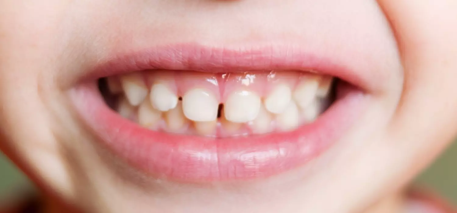
This condition can affect both primary and permanent teeth. It may affect one or more teeth, being more common in the first deciduous (temporary) lower molars.
The exact cause of this alteration is not known, but the most common factors that cause dental ankylosis are:
1. Genetic factors: A birth defect that affects the proper development of dental tissues, such as the formation of the periodontal ligament or the congenital absence of primary teeth.
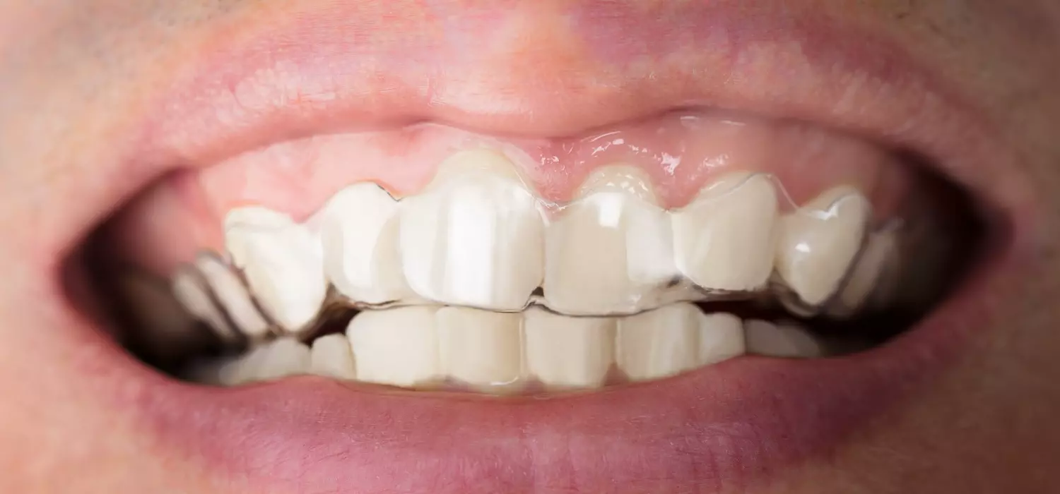
2. Habits: The consistent pressure exerted by the tongue on the teeth and persistent habits like clenching or bruxism are contributing factors. These factors are particularly relevant when considering the connection between bruxism and epilepsy.
3. Inflammatory lesions or infections: An infectious process that affects the root of the tooth and the alveolar bone.
4. Dental trauma: A blow or a strong fall involving the teeth that injures the periodontal ligament or alveolar bone.
5. Early loss of deciduous teeth: When the primary teeth fall out prematurely, a thick layer of bone forms that makes it difficult for the permanent tooth to erupt.
6. Metabolic abnormalities in the bone
7. Orthodontic treatment
8. Excessive force during chewing
9. Ankylosed teeth in children
During the dental replacement process, the permanent teeth are inside the bone at the root of the primary teeth, which is why they become loose and fall out. While the root of the primary tooth is resorbed, there are periods of rest and repair. In the reparative phase, the fusion between the cementum and the alveolar bone can develop.
Signs and Symptoms of Dental Ankylosis
Dentoalveolar ankylosis is asymptomatic; that is, it does not cause pain or discomfort. The main signs of the condition are:
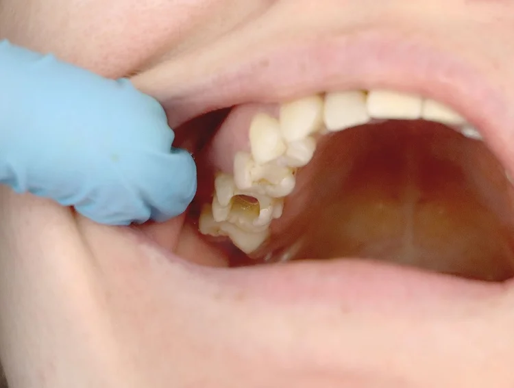
1. Teeth in infra occlusion: Affected teeth appear shorter or smaller compared to neighboring teeth as if they have not fully erupted.
2. Immobile teeth: Normally, the teeth have almost imperceptible mobility, but ankylosed teeth have no mobility at all.
3. Impacted temporary teeth: The primary tooth has not exfoliated despite having passed the indicated time to fall out.
4. Dental crowding: Misaligned and crooked teeth and a deviated dental midline can be seen in the affected arch (above or below).
5. Extruded teeth: The teeth only stop erupting when they meet the teeth of the opposing arch; for this reason, the insufficient eruption of the ankylosed tooth will generate over-eruption of the opposite tooth.
Diagnosis of Dental Ankylosis
Dental ankylosis may go unnoticed by the patient. During a visit, the dentist can make the diagnosis through a complete review that includes:
• Clinic history: The dentist will ask a series of questions to identify the medical and dental history of each patient and the possible reasons for the dentoalveolar ankylosis.
• Clinical examination: The dentist will perform a visual examination of the teeth, gums, and jawbones to identify signs of dental ankylosis.
• Radiographic examination: Periapical radiographs are essential during the diagnosis of dental ankylosis in which the union between the root and the bone and the absence of the periodontal ligament space between the 2 surfaces can be observed.
Treatment of Dental Ankylosis
Ankylosis can hinder the development of permanent teeth, so early diagnosis and intervention are essential to prevent further progression.
The treatment will depend upon the nature, position, and state of the ankylosed tooth.
1. Temporary teeth treatment
In the ankylosed teeth of children, there are two possible scenarios:
• There is a permanent tooth to replace it: The indicated treatment is the early extraction of the temporary tooth to avoid hindering the eruption of the permanent tooth. Additionally, the dentist will place a space maintainer, a device that helps preserve enough space for the final tooth to fit when it erupts.
• There is no permanent tooth to replace it (agenesia): Depending upon the age of the patient and the degree of ankylosis, the primary tooth can be kept in the mouth for life, or the dentist may extract the ankylosed tooth, maintain the space, and wait for the child to finish normal growth and development before placing a dental implant.
2. Permanent teeth treatment
Depending upon each case, the dentist can suggest several treatment options:
Surgery: In some cases, it is possible to perform surgery during which the bone around the tooth is removed to expose it and later repositioned with orthodontic treatment.
There are other surgical techniques for mobilizing the tooth with the bone, such as corticotomy and osteotomy, a procedure in which part of the bone is removed to allow for later orthodontic treatment.
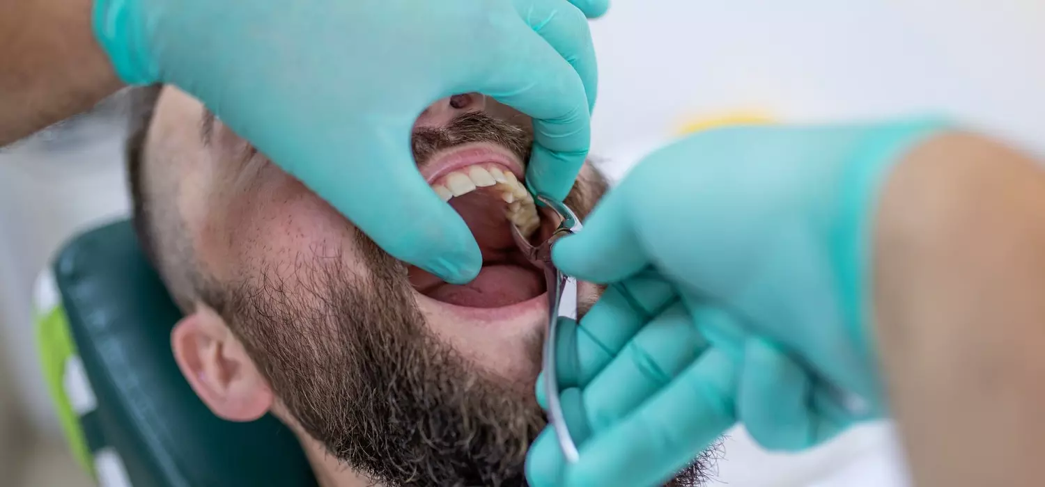
• Tooth dislocation: The tooth is firmly held with forceps and carefully moved in a gentle rocking motion, both back and forth and from side to side. Following this procedure, the periodontal ligament is reestablished to facilitate proper tooth eruption. This information is relevant for the management of tooth dislocation.
• Tooth extraction: performed when the ankylosis is very severe.
• Keep the tooth and schedule follow-up appointments: every 6 months it is important to carry out a clinical and radiographic follow-up to observe the evolution of the ankylosed tooth.
If a tooth is extracted, it must be replaced, the options are:
• Orthodontics: close the space by moving the other teeth with braces.
• Autotransplante: extract a less visible tooth and position it in the space of the missing tooth.
• Rehabilitation: placement of a dental implant, fixed or removable prostheses.
Can Dental Ankylosis be Prevented?

The condition is caused by different conditions some are not avoidable such as genetics. However, if you can prevent it, consider these tips:
• Maintain good oral hygiene: practice adequate oral hygiene techniques and use all the cleaning supplies such as a good brush, dental floss, and fluoride toothpaste. It will prevent the appearance of cavities and infections, and perhaps the loss of teeth.
• Avoid dental trauma: although it is not always possible, especially in children, it is important that when falling, they place their hands to protect their teeth from a blow.
In case of dental trauma, it is advisable to consult a dentist to determine the risk of dental ankylosis and schedule the appropriate treatment early on.
Dental ankylosis is a rare condition that alters the eruption of the teeth since their roots are attached directly to the bone, preventing them from moving naturally. The condition can occur in children and adults, both in temporary and permanent teeth. The success of the treatment will largely depend upon early diagnosis.
The recommendation is to schedule visits to the dentist at least every 6 months from 2 years of age onwards, to maintain control of each person’s dental and oral health status.
Frequently Asked Questions
Do ankylosed teeth need to be removed?
Depending upon each case, the dentist can suggest several treatment options:
Surgery during which the bone around the tooth is removed to expose it and later reposition it with orthodontic treatment, Tooth dislocation, Tooth, Keep the tooth and schedule follow-up appointments.
What does it mean if a tooth is ankylosed?
Dental ankylosis, also known as dentoalveolar ankylosis, is a condition in which the tooth is fused with the bone that supports it and thus remains immovable.
How Rare is Ankylosed Tooth?
Ankylosis is classified as a rare condition. Based on data from the International Organization of Scientific Research’s Journal of Dental and Medical Sciences (IOSR-JDMS), the prevalence of ankylosis varies between 1.3 and 14.3 percent of the population. The condition tends to occur more frequently among siblings and is somewhat more prevalent in females.
What teeth are most ankylosed?
The incidence of ankylosed teeth ranges from 1.3% to 38.5%. The mandibular first primary molars are the teeth most commonly affected, followed by the second mandibular primary molars and the maxillary primary molars.
Is it possible to move an ankylosed tooth?
Orthodontic extrusion is a specialized method used to reposition an ankylosed tooth. This technique involves the application of controlled forces to gradually pull the ankylosed tooth out of the bone and move it to its proper position over time.
Share:
References
1. Ankylosis of teeth – About the Disease. (s.f) Genetic and Rare Diseases Information Center. https://rarediseases.info.nih.gov/diseases/701/ankylosis-of-teeth/diagnosis
2. Chang, H. Y., Chang, Y., & Chen, H. (2010). Treatment of a severely ankylosed central incisor and a missing lateral incisor by distraction osteogenesis and orthodontic treatment. American Journal of Orthodontics and Dentofacial Orthopedics, 138(6), 829-838.
3. De Souza , R. F. , Travess , H. C. , Newton , T. , & Marchesan , M. A. (2015).Interventions for treating traumatised ankylosed permanent front teeth. The Cochrane library. https://www.cochranelibrary.com/cdsr/doi/10.1002/14651858.CD007820.pub3/full
4. Hadi , A. , Marius , C. , Avi , S. , Mariel , W. , & Galit , B. (2018).Ankylosed permanent teeth: incidence, etiology and guidelines for clinical management. Medical and dental research, 1(1). https://doi.org/10.15761/mdr.1000101
5. Marek, I. (May 23, 2022). Central Incisor Ankylosis in Children & Adolescents: Extraction, Distraction or Autotransplantation? American Association of Orthodontists. https://education.aaoinfo.org/aaoinfo/sessions/12107/view
6. Stanford, N. (2010). Treatment of ankylosed permanent teeth. Evidence-based Dentistry, 11(2), 44. https://doi.org/10.1038/sj.ebd.6400718
-
Nayibe Cubillos M. [Author]
Pharmaceutical Chemestry |Pharmaceutical Process Management | Pharmaceutical Care | Pharmaceutical Services Audit | Pharmaceutical Services Process Consulting | Content Project Manager | SEO Knowledge | Content Writer | Leadership | Scrum Master
View all posts
A healthcare writer with a solid background in pharmaceutical chemistry and a thorough understanding of Colombian regulatory processes and comprehensive sector management, she has significant experience coordinating and leading multidisciplina...

