Last Updated on: 27th December 2025, 08:25 am
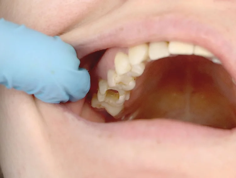
Enamel hypoplasia is a defect in the formation of the outermost layer of the tooth, and it has several implications for oral health. Many patients with dental hypoplasia consult a professional because it can severely affect their aesthetics, and therefore, their self-esteem. The good news is that there are various solutions and enamel hypoplasia treatments.
What is Tooth Enamel?
It is made up largely of the hardest mineral in the human body, called hydroxyapatite. This mineral is also an important component of bones.
What is Enamel Hypoplasia?
Enamel is the most superficial and hardest layer of the teeth. On the other hand, the word hypoplasia refers to cases in which some tissue or an organ does not fully develop.
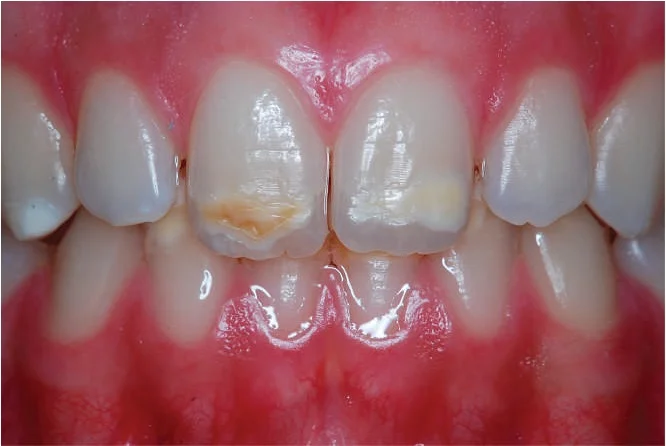
So, when talking about enamel hypoplasia, refers to a defect in which the teeth have a lower-than-normal amount of enamel. This leaves areas with a very thin enamel or the absence of it. Depending upon the cause, this anomaly can occur in one or several teeth. To learn more about enamel hypoplasia, please check out this related article.
What are the Signs and Symptoms?
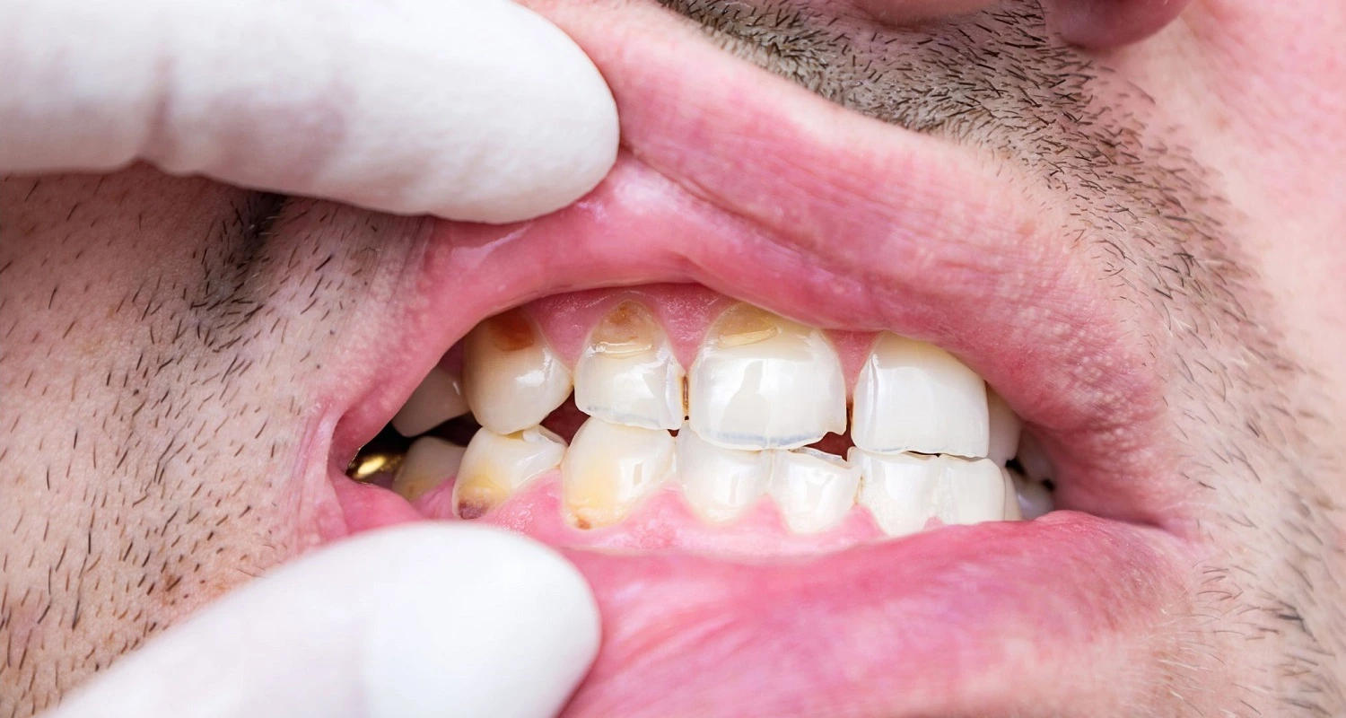
• Teeth with a stained appearance
• Changes in tooth structure
• Tooth sensitivity
• teeth more susceptible to caries, wear, and dental erosion
What Causes Enamel Hypoplasia?
Enamel hypoplasia can be caused by many factors. For example, genetic defects that cannot be prevented, or others acquired before, during, or after birth. On many occasions, the causes are not obvious. The best-known are listed below:
1. Genetics: Various hereditary syndromes affect tissues such as the skin, hair, nails, and also the teeth, generating enamel hypoplasia. Likewise, some hereditary disorders only affect the formation of dental enamel, such as amelogenesis imperfecta.
2. Metabolism affectation: An incorrect absorption of calcium and phosphorus minerals in the intestine.
3. Drugs: Ingestion of some antibiotics such as tetracyclines taken by the pregnant mother or child, during teeth formation.
4. Chemical products: Exposure to lead paint or accidental ingestion of this metal.
5. Nutritional deficiencies: Derived from alterations in metabolism or low-quality food.
6. Radiation: Product of radiotherapy treatments in the head and neck area or excessively frequent X-rays taken during the dental formation stage.
7. Trauma: Some procedures performed on newborn babies when they have respiratory distress, such as laryngoscopy, can affect tooth formation by hurting the gum as well as blows received from falls.
8. Factors associated with premature birth: Disadvantages in the functioning of the respiratory, cardiovascular, digestive, and renal systems. When anemia occurs, it can alter the formation of dental enamel.
9. Infections: When infection occurs in temporary teeth, it can damage tooth formation. Likewise, episodes of a high fever can affect the development of dental enamel.
Effects During Pregnancy
• Maternal smoking
• Vitamin D deficiency
• Congenital syphilis
Who might have Enamel Hypoplasia?
Several conditions increase the risk of presenting enamel hypoplasia:
• Premature birth
• Cerebral palsy
• Ectodermal dysplasia: the abnormal development of skin, nails, hair, and teeth.
• Family history of amelogenesis imperfecta.
• Diseases that hinder the intestinal absorption of minerals.
What care does a Patient with Enamel Hypoplasia Require?
Carrying out preventive activities and an early diagnosis of enamel hypoplasia is essential for the proper management of the condition.
For this reason, children prone to hypoplasia must be evaluated from the moment the teeth appear to adopt preventive strategies and avoid complications. Here are some basic care recommendations:
• Adopt strict oral hygiene habits, using a soft-bristled toothbrush, fluoride toothpaste, dental floss, fluoride, and alcohol-free dental rinses.
• Visit the dentist every 3 months to prevent and detect complications promptly.
• Avoid acidic drinks and foods that can cause dental erosion.
• Do not bite on objects such as pens or tools, as this could accelerate tooth wear.
Possible Complications of Enamel Hypoplasia
If enamel hypoplasia is not treated promptly and controlled over time, it can lead to some complications:
• Caries
• Increased deterioration of composite cures
• Dental fractures
• Pain
• Infection
Which Enamel Hypoplasia Treatment should you try?
The treatment of enamel hypoplasia will be focused on:
• Improving the appearance of the smile
• Preserving teeth
• Preventing complications
Depending upon the complexity of the hypoplasia, the dentist may suggest and adopt one or more of the following treatment options:
Application of fluoride or casein phosphopeptide: These substances remineralize and strengthen teeth, helping to prevent tooth decay while treating it in its early stages.
Teeth whitening: B This lightens the color of the teeth and helps to make up for the difference in color between the areas where there is no enamel and where it is less noticeable. Teeth whitening enables all the teeth to share a uniform color.
The shade used by each person will depend upon the nucleus of the tooth, its transparency, and the enamel.
However, during tooth formation, we are exposed to a series of factors that can modify the color of the teeth; in this case, due to dental hypoplasia. Aesthetic dental treatment is widely used to correct this problem.
Microabrasion: This is done by polishing with a rubber device on a rotary motor, using an abrasive paste. The dentist abrades the surface of the tooth, removing irregularities, and leaving a more uniform surface. This improves the appearance of the teeth.
Subsequently, the polished tooth layer will be filled with a composite. A dental microabrasion technique based on hydrochloric acid can be carried out without the need for rotary instruments.
Resin restorations: These are used to rebuild missing enamel and mask shape and color defects, achieving aesthetics with barely perceptible signs.
Veneers: The hypoplasia is severe, this may be a recommended solution. They are small, thin sheets of ceramic or composite (resin) that line the front of the tooth, covering imperfections and protecting the tooth. They are ideal for correcting both aesthetic and functional problems. They must be custom-made for each patient.
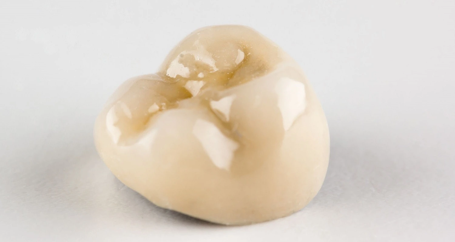
Crowns: These are indicated to treat severe cases of hypoplasia; like veneers, they are restorations that cover and protect the entire tooth on all surfaces, thus avoiding the formation of cavities and fractures as well as improving aesthetics.
They can be made of metal, ceramic, zirconium, or a combination of both materials. Of note, they are very durable. They are usually used in cases of severe enamel hypoplasia, especially in the permanent molars that erupt when children are only 6 years old.
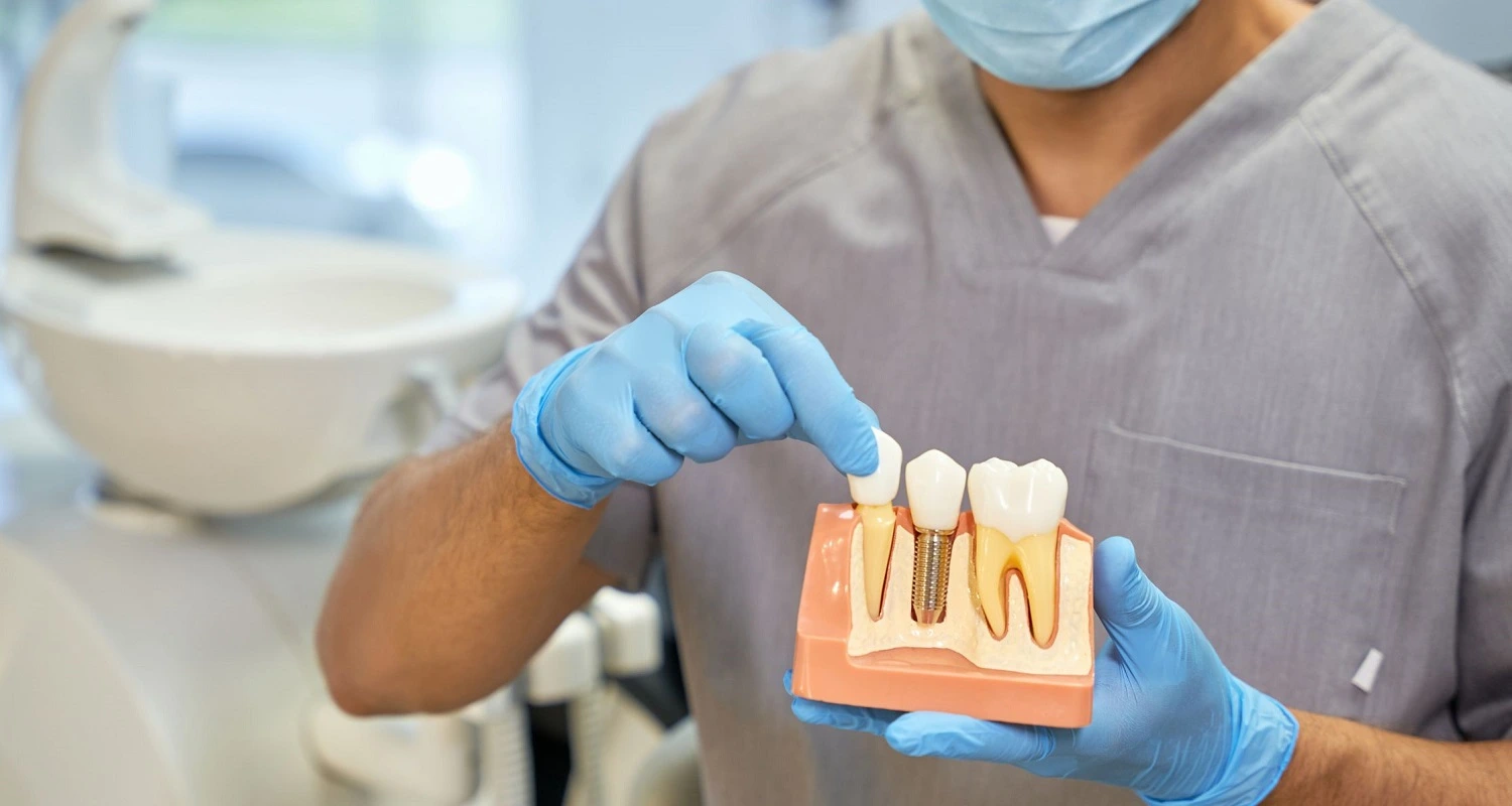
Dental implant or bridge: When this condition is severe and it is not possible to maintain the tooth, it must be removed and replaced by a dental implant. This treatment consists of replacing the roots of the teeth with bolts fixed to the jawbone and placing an artificial crown or similar piece on the superficial part of the natural tooth. This method allows for the recovery of functionality and preserves aesthetics.
Dental implant or bridge
When this condition is severe and it is not possible to maintain the tooth, it must be removed and replaced by a dental implant. This treatment consists of replacing the roots of the teeth with bolts fixed to the jawbone and placing an artificial crown or similar piece on the superficial part of the natural tooth. This method allows for the recovery of functionality and preserves aesthetics. If you would like to learn more about dental implants, you can explore this related article.
Frequently Asked Questions
What is hereditary enamel hypoplasia?
Hereditary enamel hypoplasia is a genetic defect affecting enamel formation, often seen in conditions like amelogenesis imperfecta.
What is enamel hypoplasia, and how does it affect your teeth?
Enamel hypoplasia is a defect where the enamel is thin or missing, leading to sensitivity, staining, structural changes, and a higher risk of cavities and wear.
What causes enamel hypoplasia?
Enamel hypoplasia can be caused by genetic defects, metabolic issues, certain medications, exposure to chemicals, nutritional deficiencies, radiation, trauma, premature birth, and infections. Factors during pregnancy, such as maternal smoking and vitamin D deficiency, can also contribute.
How is enamel hypoplasia treated?
Treatment includes fluoride applications, teeth whitening, microabrasion, resin restorations, veneers, crowns, and in severe cases, dental implants or bridges.
What care is required for patients with enamel hypoplasia?
Patients with enamel hypoplasia should practice strict oral hygiene using a soft-bristled toothbrush and fluoride toothpaste, avoid acidic foods and drinks, and visit the dentist every three months for monitoring and prevention of complications.
Share:
References
1. Seow, W. K. (Oct 27, 2014). Developmental defects of enamel and dentine: challenges for basic science research and clinical management. Australian dental journal, 59, 143-154. https://onlinelibrary.wiley.com/doi/10.1111/adj.12104
2. Lin, J., DMD. (Oct 25, 2022). What Is Enamel Hypoplasia? Hurst Pediatric Dentistry. https://hurstpediatricdentistry.com/2021/03/19/what-is-enamel-hypoplasia/
3. Pietrangelo, A. (Mar 13, 2018).Enamel Hypoplasia. Healthline. https://www.healthline.com/health/enamel-hypoplasia
4. Peláez, J. (Jan 13, 2021).If I have dental hypoplasia, how do I fix the loss of enamel? Ferrus&Bratos. https://www.clinicaferrusbratos.com/estetica-dental/hipoplasia-dental/
5. Elcock, C., Lath, D. L., Luty, J. D., Gallagher, M. G., Abdellatif, A., Bäckman, B., & Brook, A. H. (2006). The new Enamel Defects Index: testing and expansion. European journal of oral sciences, 114 Suppl 1, 35–379. https://onlinelibrary.wiley.com/doi/10.1111/j.1600-0722.2006.00294.x
6. Oliveira , A. , Felinto , L. T. , Francisconi-Dos-Rios , L. F. , Moi , G. P. , & Nahsan , F. P. S. (Feb, 2020). Dental Bleaching, Microabrasion, and Resin Infiltration: A Case Report of Minimally Invasive Treatment of Enamel Hypoplasia.The International journal of prosthodontics, 33(1), 105–110. http://quintpub.com/journals/ijp/full_txt_pdf_alert.php?article_id=19967
-
Nayibe Cubillos M. [Author]
Pharmaceutical Chemestry |Pharmaceutical Process Management | Pharmaceutical Care | Pharmaceutical Services Audit | Pharmaceutical Services Process Consulting | Content Project Manager | SEO Knowledge | Content Writer | Leadership | Scrum Master
View all posts
A healthcare writer with a solid background in pharmaceutical chemistry and a thorough understanding of Colombian regulatory processes and comprehensive sector management, she has significant experience coordinating and leading multidisciplina...


