Last Updated on: 27th December 2025, 05:58 am
The accumulation of fluoride in the teeth is called dental fluorosis, a condition that affects their structure and appearance. Fluoride is a naturally occurring mineral, commonly found in water.
It offers significant benefits to teeth, including the strengthening of tooth enamel and a reduction in the likelihood of cavities, among others. However, excessive fluoride consumption can lead to health issues, affecting both bones and teeth.
Why is it important to know about dental fluorosis?
Dental fluorosis first appeared at the beginning of the 20th century when a high prevalence of consultations for tooth stains later called “colorado brown stain” was typical in residents of Colorado Springs, El Paso County, in the state of Colorado.
The stains were caused by high levels of fluoride contained in the drinking water. What caught the attention of the researchers was that the people who had these spots also had a high resistance to caries. Dental fluorosis affects 1 in 4 people in the United States with mild fluorosis being the most common, 2% moderate, and 1% severe.
Dental fluorosis is not considered a disease as such, but its importance lies in the psychological effects on the person with this condition since it can be distressing and in some cases difficult to treat. It is important to regularly visit the dentist to treat these teeth on time.
What is dental fluorosis?
Dental fluorosis is a condition that occurs mainly due to excessive intake of fluoride while the teeth are being formed. Temporary teeth form during gestation while permanent teeth form between 3 to 8 years of age. Fluoride mainly affects the appearance of teeth with stains ranging from white to brown. It also affects the structure of the enamel during its formation.
Generally, the enamel is shiny and smooth, but when there is fluorosis the enamel can present an irregular appearance. The spots can be very marked or light. This condition can affect both temporary teeth and permanent teeth.
Most cases of dental fluorosis are mild. There is no pain and it does not affect the functioning of the teeth or negatively affect the quality of life. However, there are cases where the degree of dental fluorosis is severe and can affect aesthetics as well as the function of the tooth.
At this point, it is common to present pain or some type of sensitivity. Dental fluorosis is diagnosed at an early age and is very rarely diagnosed in adulthood.
Fluorosis in deciduous teeth
The formation of temporary teeth occurs during intrauterine life and continues until approximately 3 or 4 years of age. Considering that dental fluorosis appears due to the interaction of fluoride with the tooth during enamel formation, it has been found that fluorosis in deciduous teeth is caused by exposure to fluoride after birth.
The diagnosis of fluorosis in primary teeth is not easy to make since, statistically, the most affected teeth are the molars.
Fluorosis in permanent teeth
In the case of permanent teeth, which are formed between the ages of 3 and approximately 8 years, it has been shown that fluorosis is due to the interaction of fluoride with the forming enamel, depending upon the degree of exposure.
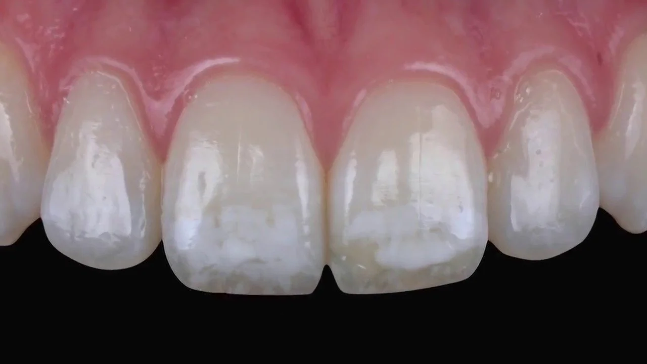
The interaction of fluoride with the tooth in formation results in spots with a whitish appearance, or brown in color with a corresponding weakening of the tooth structure.
What causes dental fluorosis?
Fluorosis is mainly caused by fluoride intake over a long period of time, precisely during the period of tooth formation. Therefore, there is a risk of fluorosis from fluoride consumption before 8 years of age. The severity of the condition depends upon the amount of fluoride, the time of consumption, and the moment it is first ingested.
Other causes of fluorosis include:
• Consumption of juices fortified with fluoride
• Fluoride fortified soft drinks
• High levels of fluoride in the local drinking water
What does dental fluorosis look like?
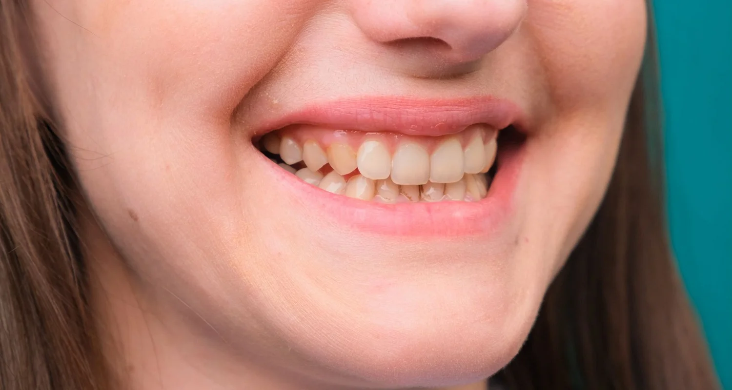
The only symptom of dental fluorosis is a discoloring of the teeth. Discoloration will vary depending upon severity. In mild fluorosis, scattered and almost imperceptible white spots are seen. To detect it, you must have special training, i.e. dentists.
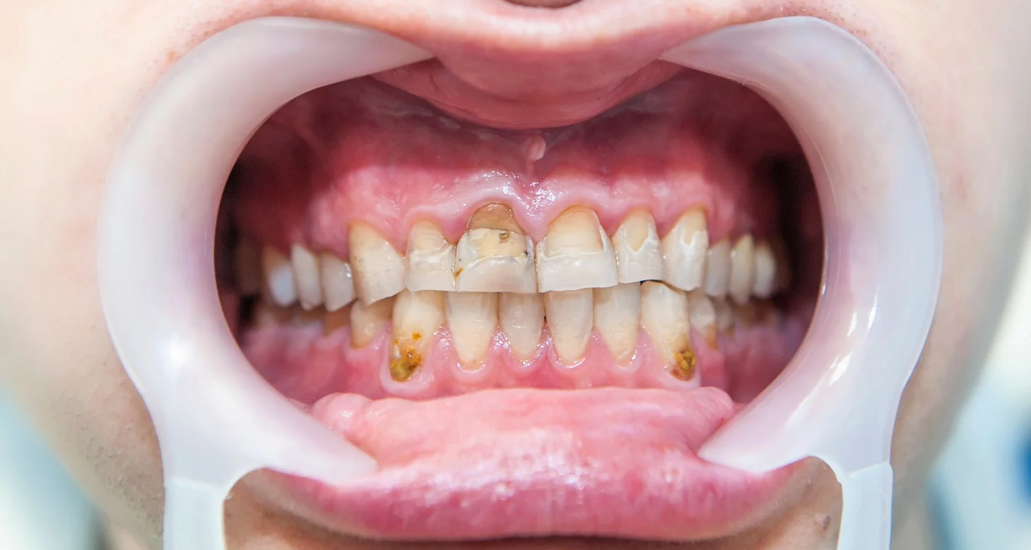
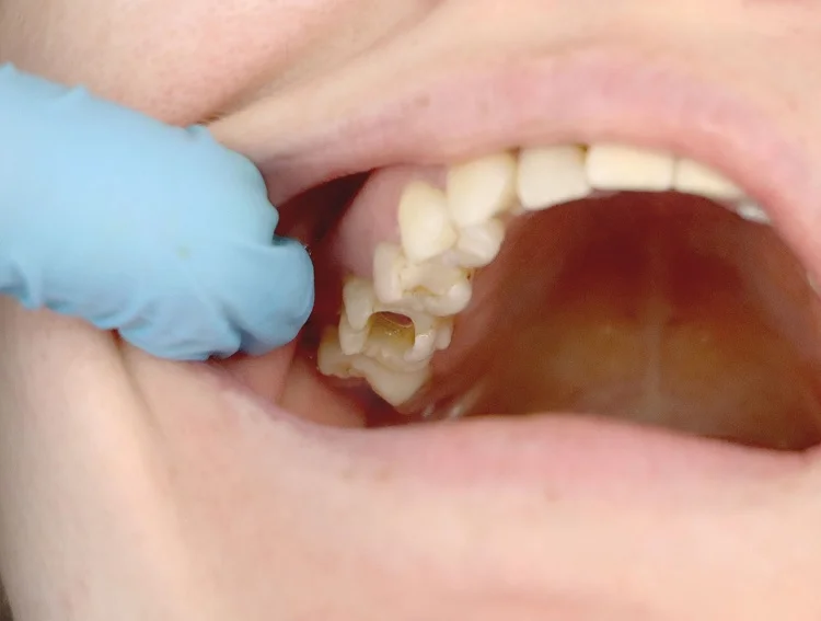
In the most severe cases, the teeth have large white or brown spots and the enamel surface is corrugated. In some cases, holes in the form of craters are observed.
What is the classification of dental fluorosis?
Dental fluorosis is classified depending on the severity:
• Questionable: White spots or spots are observed, mainly on the edges of the incisor teeth and the edge of the molars.
• Very mild: Small white, opaque areas covering less than 25% of the tooth surface are observed.
• Light: White, opaque areas covering less than 50% of the tooth surface are observed.
• Moderate: All tooth surfaces are affected and wear is also observed on the edges of the teeth and molars. The spots can be white or brown.
• Severe: All surfaces of the teeth are affected, wear is observed in the structure of the teeth along with yellow or brown holes.
Can dental fluorosis be prevented?
The risk of developing dental fluorosis is up to 8 years; to avoid its appearance you can follow the following recommendations:
Babies up to 3 years
• Exclusive breastfeeding until approximately 6 months, then incorporate solid foods and supplement with breast milk until 12 months.
• If you cannot breastfeed your baby, it is best to consult your doctor to determine the best formula.
• Brush the child’s teeth from the moment they begin to erupt, preferably two times a day.
• Use the proper amount of toothpaste, preferably no larger than the size of a grain of rice, or use only water.
Children from 3 to 8 years old
• Brushing at least 2 times a day.
• Use fluoride toothpaste sparingly: no larger than the size of a pea.
• Prevent children from swallowing the toothpaste.
• Do not use fluoride rinses on children under 6 years of age unless directed by a dentist.
Fluoride Supplements
Fluoride dietary supplements can only be used if the doctor or dentist indicates it. These supplements are recommended for children from 6 months to 16 years at a high risk of developing cavities or who live in areas that do not have fluoridated water.
Drinking water
• Under the Safe Drinking Water Act, the United States Environmental Protection Agency must notify the public if water exceeds 2.0 mg/L or parts per million. If this amount is exceeded, people must find a way to reduce it and look for an alternative to reduce the risk of fluorosis.
• If you have a well or private water supply, it is recommended to analyze the fluoride level at least once a year.
• Inform the dentist of the type of water supply as this could determine whether or not a fluoride supplement is required in the diet.
Is there treatment for dental fluorosis?
In many cases, if there is treatment, however, the severity of the condition must be determined for successful treatment. Mild dental fluorosis does not require treatment. Teeth with moderate or severe fluorosis can improve their appearance with different techniques used to mask stains rather than remove them:
• Teeth whitening: This technique is used to remove stains and make teeth whiter; however, over time it can worsen the appearance of fluorosis. If you’d like to learn more about teeth whitening, you can check out our article about it.
• Veneers: These cover the entire face of the teeth and can be made of resin or ceramic, with the latter being the most aesthetic and durable.
• Microabrasion: This technique is used to remove enamel stains. It is only recommended in mild fluorosis or with white spots that are not very pronounced.
Frequently Asked Questions
What causes a mottled appearance of enamel?
A mottled appearance of teeth, also referred to as dental fluorosis, can result from excessive fluoride intake during tooth development. This condition can arise from sources like drinking water, fluoride supplements, or certain dental products, leading to white or brown stains and uneven tooth coloration.
What causes white mottling on teeth?
White spot lesions, which indicate early tooth decay, can be caused by various factors. These include fluorosis (excessive fluoride exposure), enamel hypoplasia (thinner enamel development), enamel demineralization, a low calcium diet, and poor oral hygiene.
What is the difference between mottled enamel and fluorosis?
Mottled enamel may show yellow or brown discoloration and can have multiple pitted white-brown lesions that resemble cavities, often referred to as “mottled teeth.” In contrast, fluorosis does not initially cause discoloration. When affected permanent teeth first erupt, they are not discolored.
Can teeth whitening fix fluorosis?
Fluorosis, characterized by ribboning and discoloration of teeth due to excessive fluoride exposure, is particularly common in individuals born before 1980. This condition is challenging to treat because it does not respond effectively to topical teeth whitening methods.
How to prevent dental fluorosis?
To prevent fluorosis, monitor children to ensure they do not swallow fluoride products such as toothpaste. Supervise their brushing to minimize the amount of toothpaste they ingest.
Share:
References
1. Ansorge, R. (September 07, 2021).WebMD. Retrieved from Fluorosis Overview: https://www.webmd.com/children/fluorosis-symptoms-causes-treatments
2. American dental association (January 1, 2023).american dental association. Retrieved from Fluorosis: https://www.mouthhealthy.org/all-topics-a-z/fluorosis/
3. Campaign for dental health. (January 1, 2023).campaign for dental health. Retrieved from FACTS ABOUT FLUOROSIS: https://ilikemyteeth.org/wp-content/uploads/2014/10/FluorosisFactsforHealthProfessionals.pdf
4. Danitza Pecarevic, C.G.-L. (01 August 2022).International journal of interdisciplinary dentistry. Obtenido de Aesthetic management of dental fluorosis: Microabrasion, resin infiltration and external bleaching.: https://www.scielo.cl/scielo.php?pid=S2452-55882022000200157&script=sci_arttext
5. Farith Gonzalez Martinez, KM (October 01, 2012).Clinical Journal of Family Medicine. Obtenido de Family Factors associated with Dental Fluorosis prevalence in school children Cartagena, Colombia: https://scielo.isciii.es/scielo.php?script=sci_arttext&pid=S1699-695X2012000300006
6. J Covaleda Rodriguez, A. T.-R. (December 5, 2021).Advances in Odontostomatology. Obtenido de Minimally invasive clinical approach of dental fluorosis in stages of TF1 to TF5. Systematic review: https://scielo.isciii.es/scielo.php?script=sci_arttext&pid=S0213-12852021000200005
7. Jessica Patrícia Cavalheiro, D.G.-P. (October 17, 2017).CES dentistry. Retrieved from Clinical aspects of dental fluorosis according to histological features: a review of the Thylstrup Fejerskov Index: https://revistas.ces.edu.co/index.php/odontologia/issue/view/257
-
Nayibe Cubillos M. [Author]
Pharmaceutical Chemestry |Pharmaceutical Process Management | Pharmaceutical Care | Pharmaceutical Services Audit | Pharmaceutical Services Process Consulting | Content Project Manager | SEO Knowledge | Content Writer | Leadership | Scrum Master
View all posts
A healthcare writer with a solid background in pharmaceutical chemistry and a thorough understanding of Colombian regulatory processes and comprehensive sector management, she has significant experience coordinating and leading multidisciplina...


