Last Updated on: 30th December 2025, 09:18 am
Tooth luxation is one of the most common dental emergencies. Frequently people, both children and adults suffer some type of trauma or blow to their teeth, prompting them to go to the dentist concerned about their oral health.
This article explains dental dislocation, its signs, and symptoms, what causes it, and how it is classified.
What is tooth luxation?
The teeth are supported within the gum by two structures, the alveolar bone, and the periodontal ligament. Together they are the dental supporting tissues.
The alveolar bone is the hardest area; within this bone, each tooth is housed.
The periodontal ligament consists of soft tissue fibers that separate the root of the tooth from the bone. This ligament is elastic, that is, the fibers can stretch, shrink, and even break under certain traumatic stimuli.
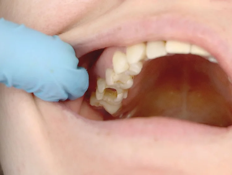
Tooth luxation is damage to the tissues that support the tooth, generally associated with trauma, falls, and blows, among others. In many cases, the dental nerve may be compromised. The teeth that most frequently suffer dental luxation are the upper central incisors, that is, the front teeth since they are more exposed to the external environment.
Signs and symptoms
The affectations of the tooth and the tissues that support it depend upon the level of severity of the trauma suffered.
The most common signs and symptoms are:
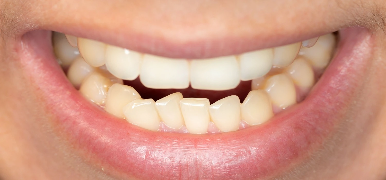
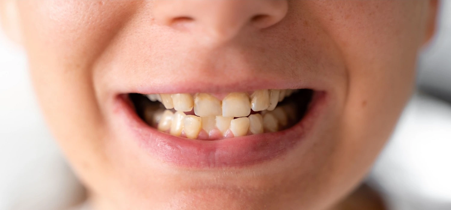
• Change of position: The affected tooth or teeth are displaced concerning their natural position: it can be inwards, outwards, or to the sides.
• Bleeding: occurs at the gum level.
• Hypersensitivity: There is an increased sensation of cold or heat when consuming hot or cold drinks or food.
• Pain: It can be mild, moderate or severe. It occurs when chewing or touching the affected tooth.
• Tooth mobility: Loosening of the affected tooth or teeth. Read more about tooth mobility right here.
• Color change: The dislocated tooth can change color and reach darker shades, such as yellow or gray, indicating involvement with the dental nerve.
Causes of tooth luxation
Tooth dislocation occurs due to trauma, such as:
• Falls
• Hits
• Violent assault
• Car accidents
• Contact sports
Those who frequently present these traumas are children when they are learning to walk, and hit the objects around them. Also, it affects people who practice a sport such as wrestling, hockey, and boxing, among others. These athletes are recommended to use dental protectors since they can receive blows to the mouth easily that may affect the teeth, gums, and supporting tissues.
Tooth luxation in children
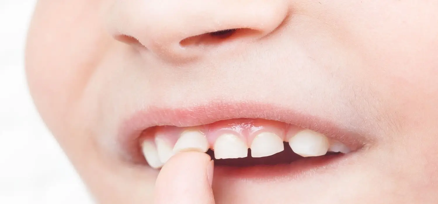
When trauma occurs at the level of the primary teeth in children, one of the most frequent consequences is permanent tooth damage.
Normally in children, the primary teeth are observed in the mouth, but at the gum level, internally, the permanent teeth are already being formed and will be maintained for the rest of the child’s life.
When a child suffers a blow to the primary teeth and luxation results, the root of that tooth likely touches and affects the teeth forming on the inside. This is important because the damage can be minimal, such as the appearance of a stain on the permanent tooth, or it can be very serious such as incurring the loss of it.
Classification types of tooth luxation
Depending upon the severity of the trauma, there are different types of dental dislocation.
• Concussion: The structures that support the tooth are affected. A W-ray will show normality, but the tooth may present pain upon chewing or receiving pressure. There is no bleeding, mobility, or change of position of the affected tooth.
• Subluxation: The affected tooth is mobile, there is bleeding at the level of the surrounding gum, and pain upon palpation. Radiographically, it will be observed that the periodontal ligament that supports the tooth is elongated. There is no change in the position of the affected tooth.
• Intrusive dislocation: The tooth moves to the bottom of the gum and becomes trapped in the gum. The tooth socket is the bone that supports the tooth; it looks shorter or may not even be seen in the mouth.
An x-ray should be taken to determine the level of tooth sinking, in which the periodontal ligament is not observed, since it is crushed against the bone. There is no mobility, but sometimes there may be bleeding, and sometimes not. Read more about irrigating wisdom tooth sockets right here.
• Extrusive dislocation: The periodontal ligament that supports the tooth is torn or stretched. There is a displacement of the tooth outward. It is observed as more elongated, and higher than the neighboring teeth, and there is mobility, pain when chewing, and bleeding at the gum level. Radiographically, the space of the periodontal ligament is observed to be increased concerning its usual shape, that is, the tooth is further separated from the bone.
• Lateral dislocation: The tooth moves forward or backward, creating a biting problem as it can interfere with the opposing teeth when chewing or closing the mouth. Bleeding at the gum level and pain are observed. The X-ray will show the increased periodontal ligament space. There is no mobility.
After a trauma, an X-ray should be taken to determine the type of dislocation, how serious it is, and if there is a fracture in the affected tooth. This will provide an adequate diagnosis and shape the best treatment plan for each case.
Treatment for dental luxation
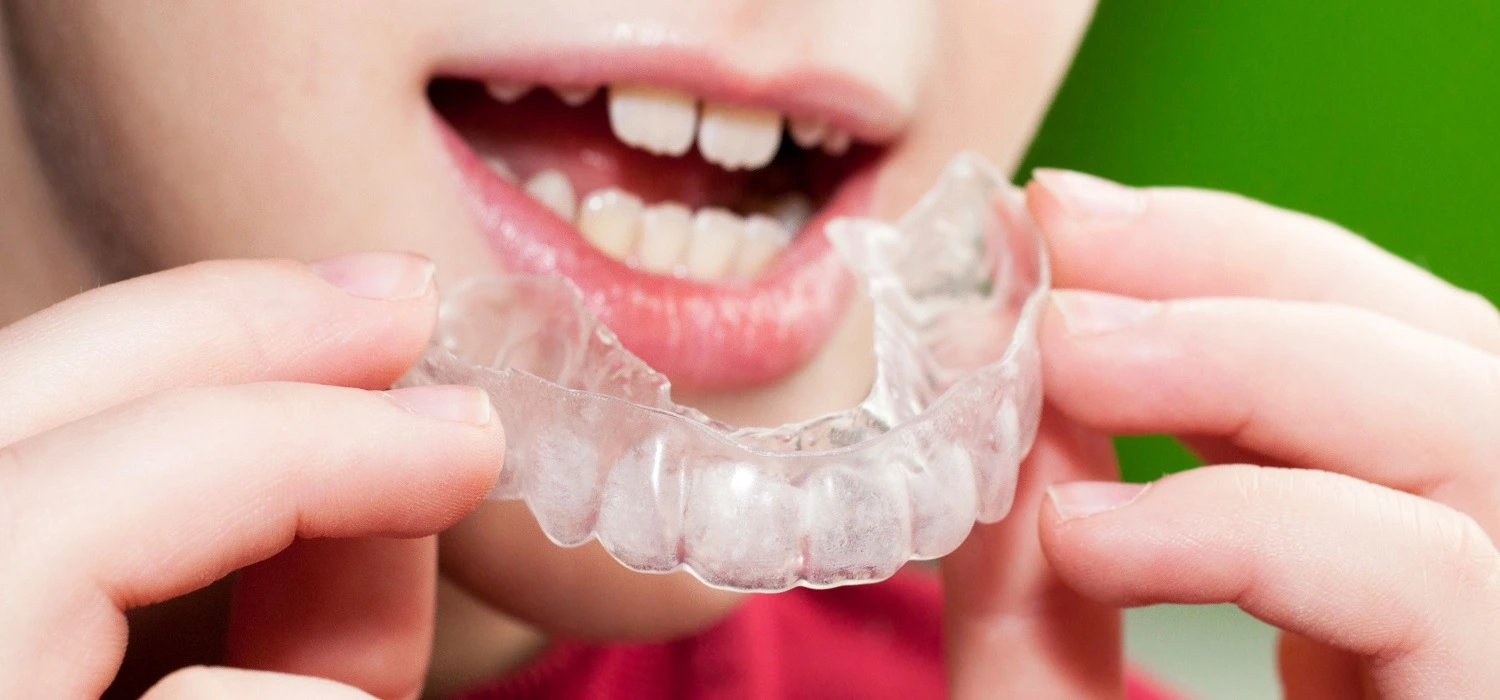
Tooth luxation, a condition where a tooth is displaced from its normal position, offers several treatment options to address the issue effectively. Repositioning involves carefully guiding the tooth back to its correct placement, while a splint can be used to stabilize the affected tooth during the healing process. Orthodontics may be employed to gradually move the tooth back to its proper position using braces or dental appliances.
In cases of internal damage, a root canal treatment may be necessary to remove injured tissue and preserve the tooth. Surgery becomes an option for more severe cases, ensuring long-term stability. Following any treatment, regular clinical and radiographic follow-ups are essential to monitor progress, assess the success of the treatment, and make any necessary adjustments.
By considering these various treatment options and maintaining diligent follow-up, dental professionals can provide optimal care and achieve favorable outcomes for patients with tooth luxation. For more detailed information about tooth luxation and treatments, you can go to our other article on the management of tooth dislocation.
Frequently Asked Questions
How long does it take for a luxated tooth to heal?
To reduce luxation, the tooth should be repositioned and secured with a flexible splint for 2 weeks, or 6-8 weeks if there is significant comminution of the alveolar bone. Follow-up examinations and radiographs should be conducted at 2 weeks, 3 months, 6 months, and 12 months. Parents and patients should be informed about the signs and symptoms of pulp necrosis.
What is the difference between tooth luxation and subluxation?
Tooth subluxation occurs when the supporting tissues of a tooth are damaged, causing the tooth to become loose without moving out of its proper position. In contrast, tooth luxation happens when the tooth not only becomes loose but also shifts out of its normal position.
What should you do with a dislocated tooth?
If you think you have a luxated tooth, visit a dentist immediately. The severity of the luxation may require urgent treatment.
How do you handle a dislodged tooth?
Holding the cleaned tooth by the crown, carefully reinsert it into the socket in the gum. Ensure that the pointed yellowish root(s) goes into the socket. This should only be done if the person is conscious. Have the person gently bite down on something soft, like a handkerchief, to keep the tooth in place.
Share:
References
1. Bourguignon , C. , Cohenca , N. , Lauridsen , E. , Flores , M.T. , O’Connell , A.C. , Day , P.F. , Tsilingaridis , G. , Abbott , P.V. , Fouad , A.F. , Hicks , L. , Andreasen , J.O. , Cehreli , Z. C. , Harlamb , S. , Kahler , B. , Oginni , A. , Semper , M. , & Levin , L. ( 2020 ).International Association of Dental Traumatology guidelines for the management of traumatic dental injuries: 1. Fractures and luxations. Dental Traumatology, 36(4), 314-330. https://onlinelibrary.wiley.com/doi/10.1111/edt.12578
2. Darley, R. M., Fernandes e Silva, C., Costa, F. D. S., Xavier, C. B., & Demarco, F. F. (2020). Complications and sequelae of concussion and subluxation in permanent teeth: A systematic review and meta‐analysis. Dental Traumatology, 36(6), 557-567. https://onlinelibrary.wiley.com/doi/10.1111/edt.12588
3. Goswami, M., Rahman, B., & Singh, S. (2020). Outcomes of luxation injuries to primary teeth-a systematic review. Journal of Oral Biology and Craniofacial Research, 10(2), 227-232. https://doi.org/10.1016/j.jobcr.2019.12.001
4. Hammel, J. M., & Fischel, J. (2019). Dental Emergencies. Emergency Medicine Clinics of North America, 37(1), 81-93. https://doi.org/10.1016/j.emc.2018.09.008
5. How do I Manage a Patient with Intrusion of a Permanent Incisor? (Updated: July 18, 2014). Canadian Dental Association. https://jcda.ca/article/e50
6. Pedrini , D. , Panzarini , S. R. , Tiveron , A. R. F. , Abreu , V. M. D. , Sonoda , C. K. , Poi , W. R. , & Brandini , D. A. (2018).Evaluation of cases of concussion and subluxation in the permanent dentition: a retrospective study. Journal of Applied Oral Science, 26(0). https://doi.org/10.1590/1678-7757-2017-0287
7. Sigurdsson, A. The treatment of traumatic dental injuries. (Summer 2014). Endodontics: Colleagues for excellence. https://www.aae.org/specialty/wp-content/uploads/sites/2/2017/06/ecfe_summer2014-final.pdf
8. Tooth Luxation: Symptoms, Causes and Treatment. (Reviewed: August 30, 2021). Cleveland Clinic. https://my.clevelandclinic.org/health/diseases/21770-tooth-luxation
-
Nayibe Cubillos M. [Author]
Pharmaceutical Chemestry |Pharmaceutical Process Management | Pharmaceutical Care | Pharmaceutical Services Audit | Pharmaceutical Services Process Consulting | Content Project Manager | SEO Knowledge | Content Writer | Leadership | Scrum Master
View all posts
A healthcare writer with a solid background in pharmaceutical chemistry and a thorough understanding of Colombian regulatory processes and comprehensive sector management, she has significant experience coordinating and leading multidisciplina...


