Last Updated on: 15th December 2025, 10:19 am
Correct dental anesthesia techniques ensure that dental procedures are conducted safely, effectively, and with minimal discomfort for patients. The techniques, which include infiltrative, troncular, intraligamentary, and submucosal anesthesia, are tailored to the specific location and type of dental procedure, ranging from simple fillings to complex extractions.
The proper use of these techniques allows dentists to work with greater precision and efficiency, reducing the potential for patient trauma. Although generally safe, dental anesthesia carries risks such as pain at the injection site, hematomas, and, in rare cases, infections or nerve damage. These potential side effects underscore the importance of administering anesthesia by skilled professionals to ensure the safety and well-being of the patient. Keep reading to learn more!
Why is it dental anesthesia?
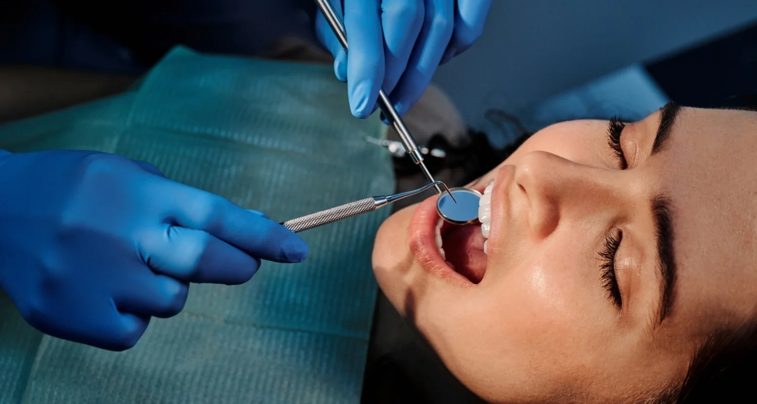
Dental anesthesia is a technique that allows dentists to block the sensitivity of a specific area of the mouth, helping to perform the required intervention in a comfortable, safe, effective, and painless way.
The person under the effects of local dental anesthesia usually relaxes a little more and avoids the tension associated with going to the dentist, since they know that they will not feel any pain.
The active ingredients present in most local anesthetics used in dentistry are compounds based on lidocaine and tetracaine. These medications work by blocking sodium channels, which are essential for initiating and transmitting sensory nerve impulses. If you want to know more about these compounds, wich are common in dental products, you can check here.
How long does the effect of local anesthesia in dentistry last?
The duration of dental anesthesia will vary based on the type of anesthetic used and the area being treated. Local anesthesia typically begins working within 10 minutes and can last between 30 and 60 minutes. For procedures like fillings or simple extractions, the numbing effect may extend for several hours after the procedure, especially for soft tissues. Factors such as protein binding and blood flow in the area also influence how long the anesthetic lasts.
With infiltrative techniques, anesthesia applied to the tooth pulp can last around 45 minutes when a vasoconstrictor like epinephrine is included. Soft tissue numbness may extend up to 1.5–2 hours. By contrast, with mild sedation, the effects wear off quickly after the procedure, allowing for a faster recovery.
Types of Local Anesthesia Used in Dentistry
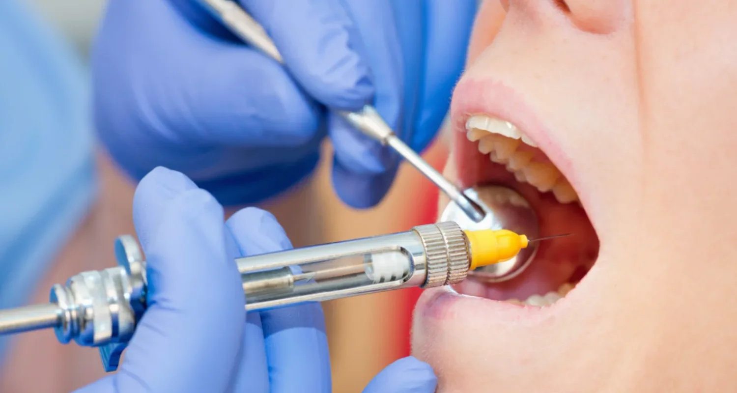
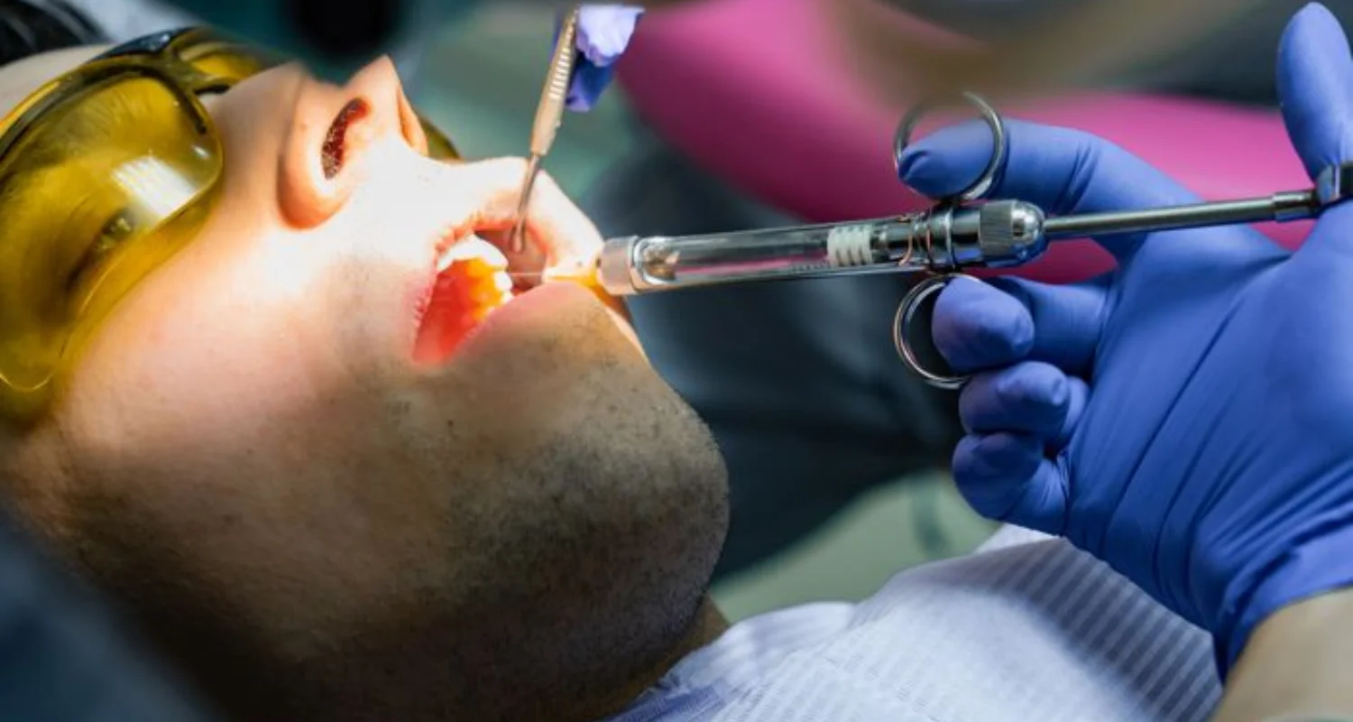
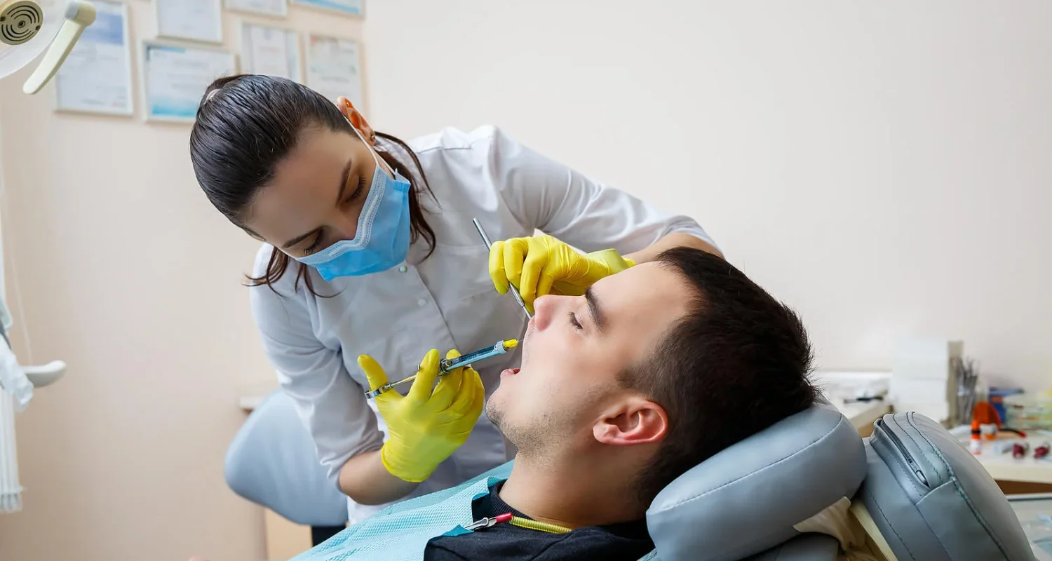
Dental anesthesia is used to numb specific areas of the mouth: a special syringe is used. In some cases, a topical anesthetic can be applied before the injection to reduce any discomfort that may arise at the time of the puncture.
This type of anesthesia is usual for the most common treatments like implants, root canals, fillings, and extractions, as well as for periodontal treatments that require a deep cleaning.
To achieve an effective anesthetic effect, various substances are used, such as lidocaine, articaine, prilocaine, bupivacaine, and mepivacaine that are administered directly to the area to be treated by injection. We find different anesthetic techniques such as the following:
Troncular
This technique, used to anesthetize the lower arch of the mouth, consists of sleeping a nerve trunk that transmits sensitivity to a large area of the oral cavity. It is commonly used on the jaw, numbing half of the mouth, including the lip and tongue on the anesthetized side. The anesthetic effect, which usually lasts at least three hours, is achieved by applying the anesthetic near the lower dental nerve, which numbs half of the arch.
Infiltrative
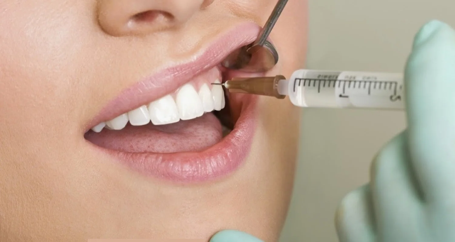
The infiltration anesthesia technique is commonly used to anesthetize the upper and lower anterior arches. It involves injecting the anesthetic directly into the area to be treated, allowing it to diffuse and reach the nerve endings. This method is particularly effective for localized procedures that do not require deep penetration into bone or other surrounding tissues.
Infiltration anesthesia is the preferred choice for dental pulp anesthesia in the upper jaw; it can also be used in the lower jaw, particularly in children for temporary teeth. While typically successful for incisors, newer formulations have shown promise for use in the molar region of the adult lower jaw. This technique provides effective localized numbness for short-term dental work.
Intraligamentary
The intraligamentary anesthesia technique is applied when other methods, such as infiltration or truncal anesthesia, are insufficient. It is commonly used for specific treatments, particularly in areas that require localized anesthesia, like endodontics. This technique can be applied to any dental unit, targeting the space between the ligament and the bone, unlike other techniques that focus on the gum.
A conventional dental syringe with a short needle or a specialized intraligamentary syringe is used. The solution diffuses from the PDL into the adjacent spongy bone under high injection pressure, providing rapid anesthesia onset, usually within 30 seconds. However, its effects are short-term, typically ranging from 10 to 45 minutes.
Submucosa
This technique consists of depositing the anesthetic solution under the mucosa, at the level of the tooth apices, without the needle coming into contact with the bone. This allows the mucosa in the infiltrated area to be anesthetized, as well as the adjacent bone and periosteum by diffusion of the solution. However, this technique does not achieve deep anesthesia of the bone, so in procedures that require direct intervention, the patient may feel some discomfort. In such cases, it is recommended to use a technique supraperiosteal or trunk, as considered by the professional.
Risks and complications of in local anesthesia in dentistry
Among the complications of anesthetic techniques we find the following:
Needle breakage
Needle fracture during dental anesthesia is rare due to the use of high-quality stainless steel needles. However, it can happen, especially with techniques like truncal anesthesia, where the needle has a longer path. Sudden movements by the patient or dentist are common causes of rupture. To minimize this risk, it’s recommended not to bend the needle, avoid inserting it fully, and use longer needles for appropriate procedures.
Symptoms of a needle fracture may include pain, swelling, restricted jaw movement (trismus), or difficulty swallowing, although some patients may not show symptoms.
Trismus
Trismus occurs when a spasm of the chewing muscles restricts the normal opening of the mouth, which should be at least 35 mm. This complication can result from trauma to the muscle or a hematoma formed during the anesthetic injection, causing limited jaw movement.
Factors like muscle penetration by the needle, the use of vasoconstrictors, or high doses of anesthesia may contribute to the development of trismus.
Conservative treatment often includes a soft diet, pain relief (analgesia), and physiotherapy to restore movement. To prevent trismus, it’s essential to use new needles with properly aligned bevels and avoid unnecessary trauma during the procedure.
Paresthesia
Paresthesia is a sensory issue that manifests as numbness, tingling, or a partial loss of sensation, often after dental anesthesia has worn off. It is commonly linked to dental procedures like wisdom tooth extractions or nerve injuries caused by instruments or high concentrations of anesthetics. Most cases resolve within 8 weeks to 6 months; but in rare instances, it can become permanent if severe nerve damage occurs.
Rare permanent cases may be due to nerve trauma from the needle or the use of anesthetics like articaine or prilocaine, highlighting the need for careful evaluation of anesthetic techniques to reduce risks.
Paralysis of the facial nerve (VII cranial nerve)
Facial paralysis in dentistry occurs when anesthetic is accidentally injected into the deep lobe of the parotid gland, affecting the branches of the facial nerve. It can be immediate, appearing a few minutes later and resolving in three hours or less, or delayed, appearing hours or days later, with a recovery time from 24 hours to several months.
Hematoma
A hematoma is caused by the rupture of a blood vessel during the injection of an anesthetic, leading to blood accumulating under the skin or in surrounding tissues. It manifests as swelling with a violet discoloration, which gradually turns bluish and fades over time. If a larger vessel is involved, symptoms like facial swelling, pain, and difficulty opening the mouth (trismus) may occur. Immediate pressure should be applied to the area to stop further bleeding; the patient should only be discharged once the bleeding is under control.
Pain during puncture and injection
When the needle is used several times in different anesthetic techniques and the same patient, it produces pain since the needle becomes blunt with use. Pain may also be noted when the anesthetic solution with vasoconstrictor is administered quickly and when the anesthetic is excessively cold.
Wrong technique
This failure is generally related to poor knowledge of head and neck anatomy. An incorrect technique is most frequently found in the lower alveolar-dentary nerve or with the truncal technique.
Side Effects of Local Anesthesia in Dentistry
Some failures that we can encounter with dental anesthesia are:
● Systemic toxicity
Systemic toxicity from dental anesthetics is rare but can happen, depending upon the injection site, dose, and type of anesthetic. Bupivacaine poses a higher risk. Symptoms can appear immediately or within 60 minutes, and interactions with other medications increase this risk. Early signs include agitation, metallic taste, confusion, drowsiness, and numbness. As toxicity worsens, symptoms like seizures, disorientation, and even cardiac arrest may occur. Proper dose management based on patient weight is crucial to avoid these effects.
● Allergic reactions
Allergic reactions to local anesthetics are relatively rare, but they can occur. Amide-type anesthetics, like lidocaine, have a lower incidence of allergic reactions, ranging from 0.1% to 1%. Some patients may react not to the anesthetic itself but to preservatives like sodium metabisulfite found in these solutions.
By contrast, ester-type anesthetics, such as benzocaine, are more commonly associated with allergic reactions, which can range from mild symptoms like itching and swelling (urticaria, rhinitis) to more severe responses such as hypotension, difficulty breathing (dyspnea), or even anaphylaxis. Dental professionals must be prepared to identify and manage these reactions effectively.
Frequently Asked Questions
Can dentists give patients too much anesthesia? What are the consequences?
Yes, it is possible for a dentist to administer too much anesthesia, although this is rare. When excessive amounts are used, it can lead to systemic toxicity, especially with agents like bupivacaine. Symptoms of toxicity may include dizziness, confusion, seizures, and, in severe cases, cardiac arrest. To avoid these effects, dentists carefully calculate the dose based on the patient’s weight and health conditions.
What type of anesthesia is used for dental work?
Several techniques are used depending on the procedure:
● Troncular anesthesia: Numbs a large area by targeting a nerve trunk, commonly for lower jaw treatments.
● Infiltrative anesthesia: Focuses on specific areas, often used in the upper and lower anterior arches.
● Intraligamentary anesthesia: Applied between the ligament and bone for precise treatments like endodontics.
● Submucosal anesthesia: Deposits anesthetic under the mucosa, typically for less invasive procedures.
These techniques ensure targeted and effective pain management during most dental work.
Can local anesthetics used in dentistry cause neurotoxicity and harm the brain?
In rare cases, local anesthetics can lead to neurotoxicity, especially if administered in high concentrations or near nerves. Neurotoxic reactions may result in paresthesia (numbness, tingling) and can affect the central nervous system, potentially causing symptoms like seizures or disorientation. Most cases resolve within weeks, but severe nerve damage could be permanent.
How long does it take dental anesthesia to wear off?
The effects of local anesthesia generally wear off within 30 minutes to a few hours, depending upon the type of anesthetic used and the area treated. Soft tissue numbness may last 1.5 to 2 hours, while more profound effects on dental pulp can last up to 45 minutes.
Share
References
1. Mathison, M., & Pepper, T. (2023, 1 junio). Local Anesthesia Techniques in Dentistry and Oral Surgery. StatPearls – NCBI Bookshelf. https://www.ncbi.nlm.nih.gov/books/NBK580480/
2. Ms, M. G. R. (2019, 13 diciembre). What You Need to Know About Dental Anesthesia. Healthline. https://www.healthline.com/health/dental-and-oral-health/dental-anesthesia#types
3. Puyuelo, C. D. (2023, 21 junio). 6 things you should know about dental anesthesia. Clínica Dental Puyuelo. https://www.clinicapuyuelo.es/en/blog/6-things-you-should-know-about-dental-anesthesia/
4. Themes, U. (2015, 12 enero). 3: Local anesthesia. Pocket Dentistry. https://pocketdentistry.com/3-local-anesthesia/
5. Marie M, Tom P,. (2023, 1 June). Local Anesthesia Techniques in Dentistry and Oral Surgery. https://www.ncbi.nlm.nih.gov/books/NBK580480/
6. Metódica. (2022, 14 septiembre). ¿Cuántos tipos de anestesia dental existen y cuál se utiliza en cada tratamiento odontológico? Centre Odontològic Els Quinze. https://els15.com/anestesia-dental/
-
Nayibe Cubillos M. [Author]
Pharmaceutical Chemestry |Pharmaceutical Process Management | Pharmaceutical Care | Pharmaceutical Services Audit | Pharmaceutical Services Process Consulting | Content Project Manager | SEO Knowledge | Content Writer | Leadership | Scrum Master
View all posts
A healthcare writer with a solid background in pharmaceutical chemistry and a thorough understanding of Colombian regulatory processes and comprehensive sector management, she has significant experience coordinating and leading multidisciplina...


